File:Rotation chamber.png
Multi-view imaging with spinning disc confocal microscopy
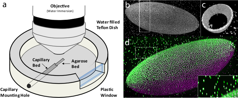
(a) Sample chamber for multi-view imaging of specimens embedded in agarose column on an upright microscope. (b) 3d reconstruction of cellular blastoderm stage Drosophila embryo imaged with 11 views on a spinning disc confocal set-up with 20x/0.5 water dipping lens. All views were deconvolved using the Huygens software. (c) Cut-out from (b) showing equal cellular resolution around the entire circumference of the embryo. (d) Superposition of maximum projection from two views (225° green and 270° magenta) highlighting their limited overlap (grey) and complementarities in specimen coverage. Inset shows overlapping point spread functions of the beads.
File history
Click on a date/time to view the file as it appeared at that time.
| Date/Time | Thumbnail | Dimensions | User | Comment | |
|---|---|---|---|---|---|
| current | 11:51, 27 May 2010 | 769 × 317 (259 KB) | Axtimwalde (talk | contribs) | Multi-view imaging with spinning disc confocal microscopy (a) Sample chamber for multi-view imaging of specimens embedded in agarose column on an upright microscope. (b) 3d reconstruction of cellular blastoderm stage Drosophila embryo imaged with 11 view |
- You cannot overwrite this file.
File usage
The following page links to this file: