File:Intensity vs Beads.jpg
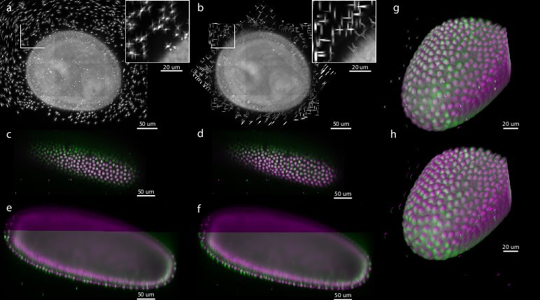
Comparison of bead-based and intensity-based multi-view reconstruction on 7-view acquisition of Drosophila embryo expressing His-YFP
(a,b) Maximum projections along the rotation axis highlight the crossing of the axially elongated bead point spread functions (inset) for (a) bead-based and (b) intensity-based registration. The raw image data for intensity-based registration were cropped to minimize the volume size. (c-f) Show cut planes through the registered specimen, where angle 0° is colored magenta and angles 45° (c,d) and 270° (e,f) are colored green to visualize the overlap of corresponding image content. Perfect overlap results in gray color. (c,e) shows the result of the bead-based registration while (d,f) shows intensity-based registration. (g,h) show 3d-renderings of the anterior portion of the embryo, colored as in (c-f). Note the increased overlap in sample intensities for the bead-based registration (g) compared to the intensity- based registration (h).
File history
Click on a date/time to view the file as it appeared at that time.
| Date/Time | Thumbnail | Dimensions | User | Comment | |
|---|---|---|---|---|---|
| current | 11:48, 27 May 2010 |  | 772 × 429 (59 KB) | Axtimwalde (talk | contribs) | Comparison of bead-based and intensity-based multi-view reconstruction on 7-view acquisition of Drosophila embryo expressing His-YFP (a,b) Maximum projections along the rotation axis highlight the crossing of the axially elongated bead point spread funct |
- You cannot overwrite this file.
File usage
The following page links to this file:
